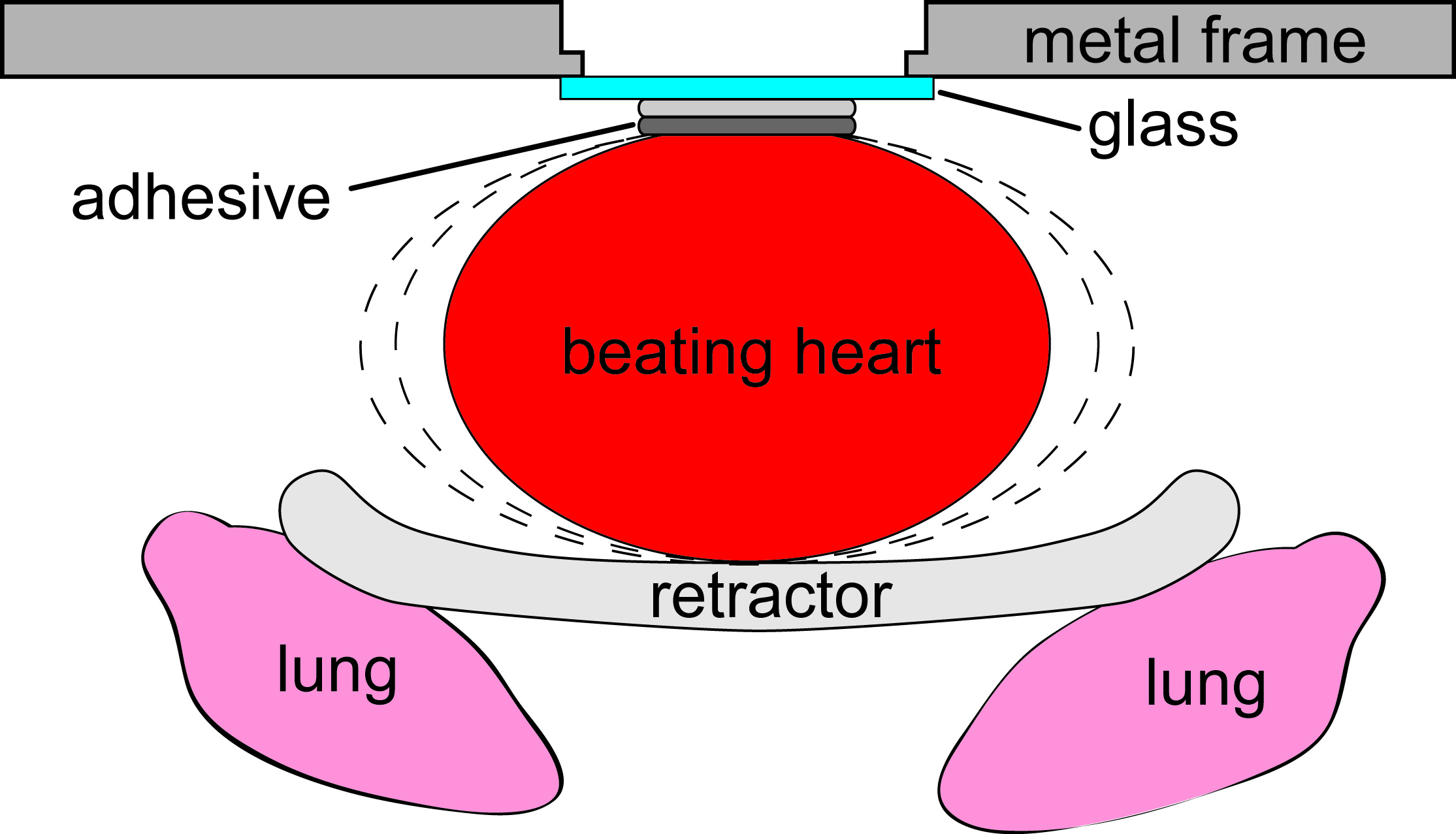
We devised a combination of surgical methods and image acquisition strategies to modify our custom-built multiphoton microscope for imaging in rodent heart in vivo. Our surgical method physically restricts the motion of a small part of the heart, while enabling most of the heart to beat freely. Image acquisition by is synchronized to the electrocardiogram.
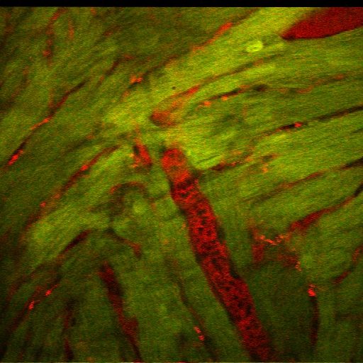
This technique enables to study the mechanisms that lead to heart attacks. We can now image capillary blood flow and the contraction of individual heart muscle cells. Using transgenic animals that express calcium-sensitive fluorescent proteins, we can also track the signals that trigger the muscle cells to contract.