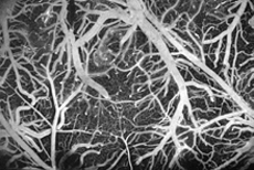
Clinical evidence shows that ischemic and hemorrhagic microvascular lesions in the brain play an important role in elderly dementia, but few effective treatment or preventative strategies exist. This deficit is due, in part, to a lack of good animal models of these small-scale strokes that would allow the progression of disease to be studied and would provide a platform for the evaluation of therapeutics.
Here, we use advanced optical techniques to induce single-vessel occlusions and hemorrhages in the cortex of live, anesthetized rodents, as a means to provide a comprehensive animal model of small-scale stroke. A tightly-focused femtosecond laser pulse is used to deposit laser energy into the endothelial cells that line a specifically targeted vessel causing an injury that triggers clotting or causes hemorrhage, but only in the targeted vessel. This technique allows any blood vessel, including individual arterioles, capillaries, and venules, in the top 0.5 mm of the cortex of a rodent to be selectively lesioned.
We also use optical techniques, such as two-photon excited fluorescence microscopy, to study the physiological consequences of these occlusions. To date, we have investigated how blood flow reroutes after the occlusion of a single vessel for clots located at different positions in the vascular hierarchy. This study has pointed to the extreme vulnerability of cortical perfusion to occlusions in penetrating arterioles and to the remarkable robustness of the cortical surface arteriole network. Ongoing work aims at determining the effect of microvascular lesions on the health and functionality of nearby brain cells.